 By Isaac Axel
The field of dentistry has evolved far beyond the traditional image of drills, manual molds, and standard bitewing X-rays. In recent years, a quiet revolution driven by advanced digital technologies has fundamentally transformed how dental professionals diagnose, treat, and monitor oral health conditions. This paradigm shift benefits both practitioners and patients. By replacing subjective assessments and invasive, uncomfortable procedures with precise, automated data, modern technology is elevating the standard of precision while directly addressing patient anxiety.
The integration of advanced engineering, data processing, and biomedical breakthroughs into dental operatories has turned what was once a highly reactive practice into a proactive, ultra-precise discipline. From the initial consultation to complex surgical interventions, digital dental ecosystems are proving that a data-driven approach yields better outcomes, minimizes recovery times, and maximizes structural longevity.
Advanced Imaging and Diagnostics
For decades, dental examinations relied heavily on two-dimensional radiographs and the physical exploration of dental surfaces using metal instruments. While these tools remain standard fundamentals, they often fail to capture micro-level changes or deep structural anomalies until a disease state has progressed significantly.
Digital Radiography
Transitioning from traditional film-based X-rays to digital imaging systems represents one of the most substantial leaps forward in everyday dental diagnostics. Digital sensors capture oral structures instantly, displaying high-resolution images on computer monitors within seconds. This immediacy allows clinicians to manipulate images by zooming, adjusting contrast, and applying filters to highlight subtle density variations in bone and enamel. From a safety perspective, digital X-rays reduce a patient’s radiation exposure by up to eighty percent compared to traditional film methods, making routine diagnostics inherently safer.
Cone Beam Computed Tomography
While standard digital X-rays provide a flat view of the teeth, Cone Beam Computed Tomography provides a comprehensive three-dimensional view of the entire maxillofacial region. This technology emits a cone-shaped X-ray beam that rotates around the patient’s head, capturing hundreds of individual images in a single rotation. Sophisticated software reconstructs these images into an accurate 3D model that details bone density, nerve pathways, soft tissues, and root canal systems.
Dentists utilize this data for highly complex procedures. For example, during dental implant placement, a clinician can evaluate the exact volume of available bone and map the precise trajectory of the implant fixture to avoid major nerve networks completely. This level of foresight eliminates guesswork, reducing the risk of surgical complications and improving the long-term success rate of restorations.
Precision Manufacturing with CAD-CAM Systems
The introduction of Computer-Aided Design and Computer-Aided Manufacturing systems has altered the logistics of restorative dentistry, particularly for dental crowns, veneers, inlays, and onlays.
Historically, getting a dental crown required a multi-step process that took several weeks. A patient had to endure a mouthful of thick, unpalatable impression putty to create a physical mold of their teeth. This mold was then packaged and shipped to an off-site dental laboratory while the patient wore a fragile temporary crown for two to three weeks. If the final restoration did not fit perfectly due to minor distortion in the physical impression material, the entire process had to be repeated.
Modern CAD-CAM workflows streamline this process into a single appointment, often referred to as same-day dentistry.
-
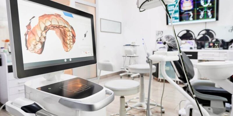
Intraoral Scanning: The dentist uses a handheld wand outfitted with advanced optical sensors to capture thousands of frames per second, creating a precise 3D digital impression of the prepared tooth.
-
Virtual Design: The digital model is loaded into specialized software where the dentist designs the exact geometry of the custom restoration, matching the natural contour and bite alignment of the surrounding teeth.
-
In-Office Milling: Once the design is finalized, the data is sent to a compact, high-precision milling machine located right inside the office. This machine carves the restoration out of a solid block of high-strength ceramic or zirconia within twenty to thirty minutes.
-
Immediate Placement: The dentist polishes, glints, and bonds the permanent crown in place during the same visit, eliminating the need for temporary restorations or follow-up appointments.
Minimally Invasive Laser Technology
Dental lasers represent another cornerstone of modern oral healthcare, offering an alternative to traditional mechanical instruments for both hard and soft tissue procedures. Operating on specific light wavelengths, lasers interact with the target tissue at a cellular level, vaporizing decayed areas or reshaping tissue with immense accuracy.
In soft tissue treatments, such as gum contouring or frenectomies, lasers cauterize blood vessels and nerve endings instantly as they cut. This results in minimal bleeding, eliminates the need for traditional sutures, and significantly dampens postoperative swelling and pain. Furthermore, the high-energy light sterilizes the surgical field on contact, drastically minimizing the incidence of secondary bacterial infections.
For hard tissue applications, lasers can selectively target dental decay without damaging the healthy, surrounding enamel structure. Because lasers generate no physical vibration or friction-induced heat, they often eliminate the localized pressure sensations that trigger dental pain. As a result, many minor cavity preparations can be completed without local anesthesia, lowering patient stress levels and reducing overall appointment times.
Artificial Intelligence and Automation
The integration of artificial intelligence into dental software suites is reshaping clinical decision-making. AI algorithms trained on massive datasets comprising millions of dental radiographs can analyze scans instantly to identify early signs of dental caries, bone loss around teeth, and periapical lesions.
Rather than replacing human expertise, AI serves as an objective, highly reliable diagnostic assistant. It flags subtle areas of demineralization that might be overlooked by a fatigued clinician, encouraging early, non-invasive interventions like fluoride therapies. Additionally, AI helps standardize diagnostic criteria across practices, increasing transparency and fostering greater patient trust in recommended treatment plans.
Frequently Asked Questions
What are smart toothbrushes, and do they actually improve daily home oral care?
Smart toothbrushes incorporate built-in pressure sensors, accelerometers, and Bluetooth connectivity to track oral hygiene routines via mobile applications. They provide real-time visual feedback detailing which areas of the mouth are being missed or brushed too forcefully, helping users modify their habits to prevent gum recession and plaque accumulation.
How does digital smile design technology help patients preview cosmetic procedures?
Digital smile design utilizes high-resolution facial photographs and 3D intraoral scans to create a virtual, highly detailed simulation of a patient’s face. Advanced software allows clinicians to alter the shape, size, color, and positioning of teeth on screen, allowing patients to visualize and approve their final cosmetic outcomes before treatment begins.
Why is silver diamine fluoride considered a major technological advance in pediatric dentistry?
Silver diamine fluoride is a non-invasive, painted-on liquid medication that stops the progression of dental cavities instantly without the need for drilling or local injections. It is a vital tool for managing decay in very young children, uncooperative pediatric patients, or individuals with special needs who cannot tolerate traditional restorative procedures.
How do modern dental hard-tissue lasers differentiate between healthy enamel and a cavity?
Hard-tissue dental lasers operate at specific light wavelengths that are highly absorbed by water and the mineral content found in decayed tooth structures. Because dental decay contains a significantly higher percentage of water than healthy, dense enamel, the laser energy targets and vaporizes the decayed cells while leaving the sound enamel intact.
What is guided implant surgery, and how does it utilize computer data?
Guided implant surgery involves taking 3D data from a cone beam CT scan and combining it with a digital impression to design a custom surgical guide. This physical template is 3D-printed and placed inside the patient’s mouth during surgery, featuring precise sleeves that lock the surgeon’s drill into the exact pre-planned depth, angle, and position.
How have modern advancements changed the materials used for orthodontic aligners?
Modern clear aligners utilize advanced, medical-grade thermoplastic materials engineered to apply continuous, highly controlled forces to teeth. These specialized plastics offer superior clarity, stain resistance, and flexibility compared to early generations, resulting in more predictable tooth movement and enhanced comfort for patients.